
Artificial Intelligence In Breast Cancer Treatment: How AI Supports Diagnosis, Planning, And Care

Imaging analysis and pathology integration in AI for breast cancer
Imaging analysis and digital pathology are among the most visible AI applications in breast cancer workflows. Imaging algorithms often process mammography, tomosynthesis, ultrasound, or MRI to highlight areas that warrant focused review. Pathology platforms commonly analyze digitized slides to quantify features such as mitotic figures, cellular density, or biomarker staining intensity. These outputs can serve as structured inputs for multidisciplinary case review, where radiologic and pathologic findings are considered together. Care teams may view algorithmic outputs as one data layer alongside human interpretation and additional testing.
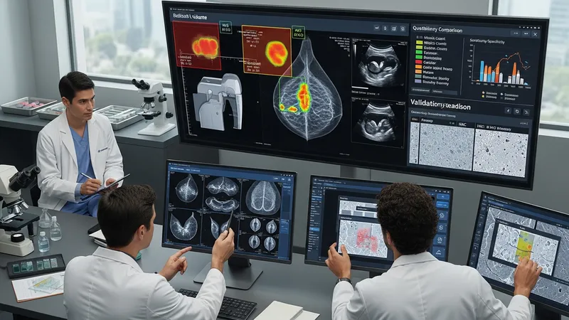
Performance differences across imaging modalities and lab processes can affect how an algorithm is used. For example, image acquisition settings, vendor equipment, and staining protocols can change input characteristics. Developers and institutions often report validation on specific scanner types or staining workflows, so implementers may run local verification studies. In research literature, reported improvements in sensitivity or specificity are typically context-dependent; therefore, local clinical validation and calibration are often described as prudent steps before routine reliance.
Interoperability between imaging systems, digital pathology viewers, and electronic records is an implementation consideration. Integration facilitates consolidated reporting and reduces manual transcription errors, but it may demand technical work such as mapping data fields and ensuring secure data transfer. Institutions sometimes prioritize staged deployments—starting with read-only or triage modes—so clinicians can compare algorithm output with standard practice before changing diagnostic pathways.
Operational governance around imaging and pathology AI generally includes labeling expected use cases, documenting performance metrics in real-world settings, and defining clinician responsibilities for review. Clinical workflows frequently specify that algorithm outputs require human confirmation and are to be interpreted in the clinical context. Reporting mechanisms may capture discordant cases to inform iterative model updates and maintain a record of decision rationale.